外国人向け【脳ドック+PET/CT がん検診】
PET/CTがん検診に頭部MRI検査による脳ドックが追加されたコースです。
MRI検査とは
MRI(=Magnetic Resonance Imaging、磁気共鳴画像診断装置)検査は、強力な磁石でできた筒の中に入り、磁気の力と電波を利用して体の臓器や血管を撮影する検査です。

がんの有無や広がり、他の臓器への転移がないかを調べる、治療の効果を判定する、治療後の再発がないかを確認するなど、さまざまな目的で行われる精密検査です。(がん情報サービス、MRI検査とはhttps://ganjoho.jp/public/dia_tre/inspection/mri.html
MRI検査は体内のあらゆる方向の断面像を詳しく見ることができますので、様々な病巣の発見に有効です。特に脳や脊椎、腹部臓器、卵巣、前立腺などの下腹部、四肢などの関節の撮影を得意とします。
X線被ばくがないので、安心して繰り返し検査することができる利点があります。
頭部MRI検査とは?
MRIを使って頭部の検査をすることを「頭部MRI検査」と呼んでいます。
頭部MRI検査は、脳の断面画像を得ることができますので、脳の異常を早期発見するのに有効です。
頭部MRI検査のメリット
✔ あらゆる方向の脳の断面像が撮影できる
✔ CT検査より解像度が高く、細かく脳の状態を評価できる
✔ 放射線を使わないため、被ばくがない
造影剤を使用することにより、脳腫瘍や血管の診断にも有効です。
※今回の検診メニューの脳ドッグは、造影剤なしの単純MRI検査です。
頭部MRI検査で発見される主な疾患
認知症や脳腫瘍、脳梗塞、脳出血等の脳血管障害
頭部MRI検査のデメリット
✔ 狭い空間で大きな機械音がする、閉所や騒音が苦手な方は苦痛を感じる可能性がある
✔ 強力な磁場が発生するため、金属類が体内に入ってある場合は、受けられない可能性がある
【脳ドック + PET/CT がん検診】
脳ドックは認知症、脳腫瘍、脳梗塞や脳出血等の脳血管障害の早期発見、予防を目的とする健康診断です。
PET/CTがん検診は、全身のがん有無を調べることが目的です。
当院の【脳ドック+PET/CTがん検診】は、頭部MRI検査とPET/CT(FDG)検査を同じ日に実施します。頭部MRI検査は、造影剤を使わない単純MRI検査です。
頭部MRI検査のみは対応しません。また、上記の検査を別日程でそれぞれ実施することも対応できませんので、あらかじめご了承ください。
こんな方におすすめ
✔ がんや脳血管障害を早期に発見したい方
✔ ご家族にがんの既往がある方
✔ 認知症や脳出血、脳梗塞など血管障害の家族歴がある方
受けられない方
✔ 心臓ペースメーカーや埋め込み型除細動器(ICD)を使用している方
✔ 極度の閉所恐怖症の方
✔ 自立歩行が困難な方
✔ ひとりでのトイレが難しい方
✔ 認知機能低下がある
✔ 1年以内のてんかん発作がある方
✔ 妊娠中、または妊娠している可能性がある方
✔ 授乳中の方
検査前にお申し出いただきたいこと
✔ 治療のため身体の中に金属が入っている(脳動脈クリップ、ステント、人工弁、人工関節など)が埋め込まれている
✔ 磁石を使った医療機器を装着している
✔ 事故などで体の中に金属片が入っている(またはその可能性がある)
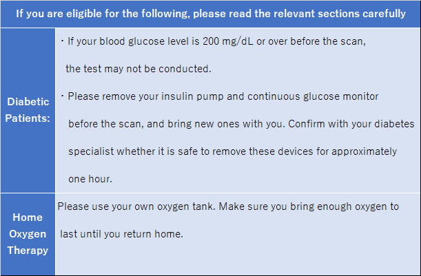
✔ インスリンポンプやグルコース測定器を使っている
✔ DIBキャップ(磁石式尿道カテーテルキャップ)を使っている
✔ ニトロのパップ剤や禁煙パッチを使っている
✔ 刺青、アートメイクがある方
該当される方は、検診申込票のチェックリストに記載の上、事前にお申し出ください。
【脳ドック + PET/CT がん検診】の流れ
受付
当日、予約票に記載された受付時間までに、総合案内にお越しください。
当院コーディネーターが放射線センター受付までご案内します。
更衣
更衣室で着替えをしていただきます。着替えがしやすい服装でご来院ください。
検査に影響を及ぼすため、以下のものはすべて取り外していただきます。
➤ 金属類:ヘアピン、鍵、メガネ、財布、アクセサリー類、ベルト、ライターなど
➤ カード:クレジットカード、キャッシュカード、定期券、交通系カードなど
➤ 電子機器:携帯電話、タブレット、時計など
➤ その他:補聴器、取り外せる入れ歯、金属が入った下着、エレキバン、ホッカイロ、
湿布、ニトロダーム、ニコチネルなど
更衣室から出る際は、ロッカーのカギ、会計用ファイルのみお持ちください。
MRI撮影
着替えが完了したら、スタッフがMRI検査室にご案内します(足元の緑ラインに沿って進みます)。
頭部MRIを20分程度撮影します。
完了したら、再びPET検査室まで戻ります。足元の白いラインに沿って進んでください。
診察
PET/CT検査前に問診と血糖値測定、検査の流れなど説明を行います。
FDG薬剤の投与
腕の静脈にFDG薬剤を注射します。
待機
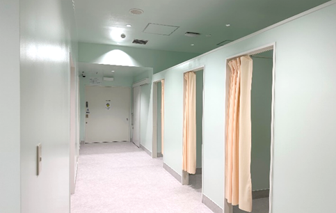
薬剤が全身に回るまで、指定の待機場所で約60分間、安静にしてお待ちいただきます。
携帯電話を見たり、本を読んだりすることもできません。
安静を保てない場合、検査を行えなくなることがございます。
検査室内、待機エリアともにミントグリーンを
基調とした穏やかな空間です。
撮影
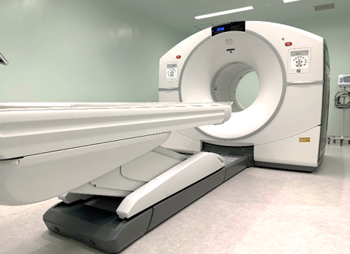
PETカメラとCTが合体したPET/CT装置で撮影します。
撮影時間は約30分間です。
PET/CT装置(DiscoveryIQ)
待機
撮影終了後、FDGの放射能が減少するまで、指定場所で約30分間お待ちいただきます。
お会計
すべての検査終了後、コーディネーターが会計窓口へご案内いたしますので、お支払いをお願いいたします。
【脳ドック + PET/CT がん検診】の注意事項
| 検査前 | 「受けられない方」に該当がある方は必ず事前に申し出てください。 |
|---|---|
| 検査前日から終了までは、激しい運動や肉体労働などはしないでください。 | |
| 糖尿病薬を服用中の方は、申込時に申込書の記載欄に薬品名をご記載ください。 | |
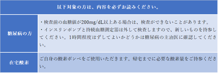
| 検査当日 | PET/CT検査のため、検査予約時間の6時間前から禁食が必要です。お水はお飲みいただけます。糖分が入った飲み物は禁止です。 |
| 糖尿病薬を服用中もしくはインスリン療法の方は、検査予約時間の6時間前から中止してください。 | |
| 金具やアクセサリー、金属をなるべく使用していない服装でご来院ください。 | |
| 化粧、マニキュア、カラーコンタクトレンズなどは使用せずご来院ください。 | |
| 日本語ができない場合は、日本語のできる方の同伴をお願いします。 | |
| 検査後 | 検査終了後、1~2時間はできるだけ人混みを避けてください。 |
| 水分は多めにとるようにしてください。 | |
| 検査した日は、男性の方も座って排尿してください。 |
Q&A
| PET/CT(FDG)検査に関する【Q&A】は、PET/CTがん検診ページをご確認ください。 | |
| Q | MRI検査はなぜ大きな音が出るのでしょうか? |
| A | 体内臓器の位置情報などを得るためには、磁場の強さや向きが変動する「傾斜磁場」が必要となります。MRI装置内部に取り付けられたコイルは、磁場を高速に切替える際の大きな力によって振動してしまいます。そのため、撮影の仕方によって「ガンガンガン」、「ピーピーピー」、「ダダダダ」といった様々な大きな音が発生するのです。(日本画像医療システム工業会企画「MRIのQ&Aより」 |
|---|---|
| Q | 頭部MRIは頭部CTとは違うのでしょうか。 |
| A |
頭部MRIは磁気を利用した検査、CTは放射線(X線)を利用した検査で、CTは被ばくが発生します。 一般的には、MRIはCTより解像度が高く、細かく脳の状態を評価できるといわれています。また、造影剤を使わずに主な血管の画像が簡単に得られます。 ただし、MRIでは骨の情報はほとんど得られません。CTは脳出血や頭蓋骨骨折の描出など骨の異常の発見には有効です。 |
| Q | MRI検査時間はどれぐらいかかるでしょうか? |
| A | 今回の脳ドッグは、頭部MRIのみで、20分ぐらいかかります。 |
| Q | 検査の間、体を動かすことはできますか? |
| A | 検査の間は、できるだけ体を動かさないで、静かに横になっていてください。 |
| Q | 検査中、気分が悪くなったらどうしますか? |
| A | 呼び出しブザーを渡しますので、気分が悪くなった場合は、遠慮せずにブザーを押してください。 |
Head MRI & PET/CT Scan
This course combines a PET/CT cancer screening with a Brain screening, which includes a head MRI scan.
What is MRI Scan?
MRI(Magnetic Resonance Imaging)is an imaging test that uses a powerful magnet and radio waves to capture detailed images of organs and blood vessels inside the body.
 It is used for various purposes, such as detecting cancer, assessing its spread, checking for metastasis, evaluating treatment effectiveness, and monitoring for recurrence after treatment.
がん情報サービス (Gan joho servise)
cancer information sarvice
MRI検査とは(What is an MRI scan ?) https://ganjoho.jp/public/dia_tre/inspection/mri.html
MRI can provide detailed cross-sectional images from multiple angles, allowing for the detection of various abnormalities. It is particularly effective in imaging the brain, spine, abdominal organs, ovaries, prostate, and joints of the limbs. Since MRI does not involve radiation exposure, it can be safely repeated as needed.
What is a Head MRI Scan?
A head MRI scan is an imaging test that examines the brain using an MRI machine.
It provides cross-sectional images of the brain, making it an effective tool for early detection of abnormalities.
Benefits of Head MRI Scans
✔ Captures cross-sectional images of the brain from multiple angles
✔ Provides higher resolution than a CT scan for detailed brain evaluation
✔ Does not use radiation, eliminating exposure risks
With the use of contrast agents, MRI can also help diagnose brain tumors and blood vessel conditions.
* The Brain Screening is done without the use of contrast agent.
Conditions That Can Be Detected
Dementia, brain tumors, cerebrovascular diseases, including cerebral infarction and hemorrhage.
Limitations of Head MRI Scans
✔ The scan takes place in a confined space and involves loud machine noise, which
may be uncomfortable for those who are claustrophobic or sensitive to noise.
✔ Due to the strong magnetic field, people with metal implants in their body may not
be eligible for the scan.
Head MRI & PET/CT Scan
A Brain Screening is a health screening aimed at the early detection and prevention of dementia, brain tumors, and cerebrovascular diseases such as stroke, including cerebral infarction and hemorrhage.
A PET/CT cancer screening is designed to check for the presence of cancer throughout the body.
At our hospital, the Head MRI + PET/CT Cancer Screening includes a head MRI scan and a PET/CT (FDG) scan, both performed on the same day. The head MRI scan is a non-contrast MRI.
Please note that we do not offer a head MRI scan alone, nor can we schedule the above tests on separate days.
Head MRI Scans are recommended for:
✔ Those who want to detect cancer or cerebrovascular disease at an early stage
✔ Those with a family history of cancer
✔ Those with a family history of dementia, cerebral hemorrhage, cerebrovascular diseases or other vascular diseases
Not suitable for Head MRI Scans
✔ Those who with a pacemaker or an implanted defibrillator
✔ Those with severe claustrophobia
✔ Those who have difficulty walking independently
✔ Those who need assistance using the toilet
✔ Those with cognitive impairment
✔ Those who have had a seizure within the past year
✔ Those who are pregnant or may be pregnant
✔ Those who are breastfeeding
Please inform us before the examination if:
✔ You have metal implants in your body for medical treatment (e.g., cerebral artery
clips, stents, artfitifical heart valves, artificial joints).
✔ You are using a medical device that contains magnets.
✔ You have, or you might have, metal fragments in your body due to an injury or
accident.
✔ You use an insulin pump or a glucose monitor.
✔ You use a DIB cap (magnetic urinary catheter cap).
✔ You are using a nitroglycerin patch or nicotine patch.
✔ You have tattoos or permanent makeup
If any of the above apply to you, please check the relevant boxes on the screening application form and inform us in advance.
Flow of Head MRI & PET/CT cancer screening
Reception
On the day of your appointment, please arrive at the Information Desk by the check-in time listed on your reservation slip. Our coordinator will guide you to the Radiology Center Reception.
Changing Clothes
After Check-in, you will need change into examination clothes in the changing room.
Please wear comfortable clothing that easy to change.
For the examination, you must remove the following items as they may affect the scan.
Metal items: Hair pins, keys, eyeglasses, wallets, accessories, belts, lighters, etc.
➤ Cards: All magnetic or IC cards such as credit / cash cards, and transit cards, etc.
➤ lectronic devices: Mobile phones, tablets, watches, etc.
➤ Other: Hearing aids, removable dentures, underwear with metal parts, electromagnetic therapy patches, disposable heat packs, medicated patches, nitroglycerin patches, nicotine patches, etc.
When leaving the changing room, bring only your locker key and payment clear file.
MRI Scan
Once you are ready, a staff member will guide you to the MRI room (follow the green line on the floor.)
The head MRI scan takes approximately 20 minutes. After completion, please return to the PET/CT examination area by following the white line.
Consultation
Before the PET/CT scan, we will conduct a medical interview, measure your blood glucose level, and provide necessary explanations.
FDG Tracer Injection
In the examination room, FDG tracer will be injected into a vein in your arm.
Resting
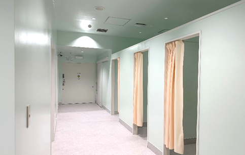
After the injection, you will need to rest in a designated waiting area for approximately 60 minutes while the tracer spreads throughout your body. During this time, you cannot use your mobile phone or read books. If you are unable to remain still, we may not be able to proceed with your scan.
Both the examination room and thewaiting area are designed with a calming mint green theme.
Scanning
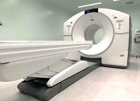
The imaging will be done using a PET/CT scanner, which combines a PET camera and a CT scanner. The scanning time is approximately 30 minutes.
PET/CT(Discovery IQ)
Waiting
After the scan, you will need to wait in a designated area for approximately 30 minutes until the radiation from the FDG in your body decreases.
Payment
After all your scans are completed, the coordinator will guide you to the Accounts desk for your payment.
Precautions for Head MRI + PET/CT Cancer Screening
| Before the Scan | If any of the conditions under “Not Eligible or Not Suitable” apply to you, please inform us in advance. |
|---|---|
| Avoid strenuous exercise or heavy physical labor from the day before the scan until it is completed. | |
| If you are taking medication for diabetes, please write the name of the medication in the designated. | |
| On the day of the Scan | For the PET/CT scan, you must fast for six hours before your scheduled appointment time. You may drink water, but beverages containing sugar are not allowed. |
| If you are taking diabetes medication or undergoing insulin therapy, stop taking it six hours before your scheduled appointment time. | |
| Please wear clothing without metal, accessories, or metallic components as much as possible. | |
| Do not wear makeup, nail polish, or colored contact lenses. | |
|
If you do not understand Japanese, please bring an interpreter with you. |
|
| After the Scan | After the scan, avoid crowded places as much as possible for one to two hours. |
| Drink plenty of fluids. | |
| On the day of the scan, men should sit while urinating. |
Q&A
For information about PET/CT (FDG) exams, please see the Q&A section on the PET/CT Cancer Screening page.
Q : Why does an MRI scan make loud noises?
A :
To obtain information about the position of internal organs, an MRI scan requires a "gradient magnetic field," which involves changes in the strength and direction of the magnetic field. The coils inside the MRI machine vibrate due to the strong forces generated when the magnetic field switches rapidly. This results in various loud sounds, such as "bang bang bang," "beep beep beep," or "da da da," depending on the scanning method.
(*Source: Japan Medical Imaging and Radiological Systems Industries Association, "MRI Q&A"*)
Q: How is a head MRI different from a head CT?
A :
A head MRI uses magnetism, while a CT scan uses radiation (X-rays), which involves exposure to radiation.
In general, MRI provides higher resolution than CT and allows for a more detailed evaluation of brain conditions. Additionally, MRI can easily capture images of major blood vessels without using contrast agents.
However, MRI does not provide much information about bones. CT is useful for detecting abnormalities such as brain hemorrhages or skull fractures.
Q : How long does an MRI scan take?
A:
This brain screening includes only a head MRI and takes about 20 minutes.
Q : Can I move my body during the scan?
A :
Please remain as still as possible and lie quietly throughout the scan.
Q: What should I do if I feel unwell during the scan?
A:
You will be given a call button. If you feel unwell, please press it without hesitation.
外国人【脑部精检+PET/CT癌症筛查体检】
此项目在PET/CT癌症筛查体检的基础上添加了头颅磁共振检查的脑部精检。
什么是磁共振(MRI)检查?
磁共振(MRI=Magnetic Resonance Imaging,核磁共振成像)检查是,进入一个由强力磁铁制成的管子中,利用磁力和无线电波拍摄人体器官和血管的影像学检查。
 磁共振检查是一项精密检查,其目的多种多样,包括检查有无癌症及其扩散程度,是否转移到其他器官,评估治疗效果,确认治疗后有没有复发等。(がん情報サービス:癌症信息服务网、MRI検査とは:什么是磁共振检查https://ganjoho.jp/public/dia_tre/inspection/mri.html)
磁共振检查可以获得体内各个方向的断层图片,检测出体内的各种病变。尤其擅长对大脑,脊椎,腹部器官,卵巢,前列腺等下腹部,及四肢等关节的成像。
由于没有核辐射,因此可以放心并重复检查是其优点。
什么是头颅磁共振检查?
头颅磁共振检查是指用核磁共振成像检查头颅。
头颅磁共振检查,可以获得头部的断层图片,对早期发现脑部异常有效。
头颅磁共振检查的优点
✔ 可以拍摄各个方向的脑部断层影像
✔ 比CT检查,图像的分辨率高,能够详细评估脑的状态
✔ 不使用放射线,没有核辐射
使用造影剂,会对脑肿瘤和血管的诊断更加有效。
※本院提供的脑部精检是不使用造影剂的磁共振检查。
头颅磁共振检查可以发现的主要疾病
痴呆症,脑肿瘤,脑梗塞,脑出血等脑血管障碍
头颅磁共振检查的弊端
✔ 对于不喜欢封闭空间或噪音的人,进入狭窄的空间伴随极大的机器噪音可能会让人感到痛苦
✔ 会产生强大的磁场,因此体内有金属材料时不能接受检查
【脑部精检+PET/CT癌症筛查体检】
脑部精检是为了预防和早期发现痴呆症,脑肿瘤,脑梗塞,脑出血等脑血管障碍。
PET/CT癌症筛查体检的目的是检查全身是否有癌症。
本院的【脑部精检+PET/CT癌症筛查体检】,同一天进行头颅磁共振和PET/CT(FDG)检查。头颅磁共振检查不使用造影剂。
不单独接受头颅磁共振检查。此外,关于上述两项检查,不能分别在不同日期进行,请悉知。
推荐给以下人群
✔ 想早期发现癌症或脑血管障碍的人
✔ 家族中有癌症病史
✔ 有痴呆症,脑出血,脑梗塞等脑血管障碍的家族病史
无法接受本体检的人
✔ 体内植入有心脏起搏器或除颤器(ICD)的人
✔ 有严重的封闭恐惧症
✔ 独立步行困难
✔ 一个人如厕困难
✔ 认知功能低下
✔ 最近一年内有过癫痫发作
✔ 孕期,或目前有怀孕的可能性
✔ 哺乳期女性
检查前须告知的事项
✔ 为治疗体内植入了金属材料(脑动脉夹,支架,人工瓣膜,人工关节等)
✔ 身上佩戴有带磁石的医疗器械
✔ 因事故体内可能残留了金属(或有残留的可能性)
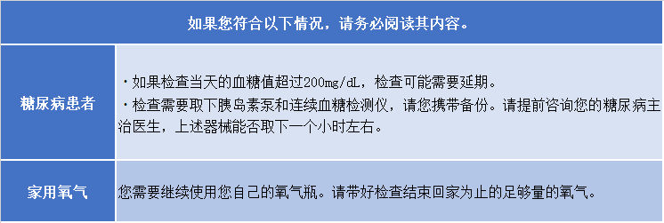
✔ 在使用胰岛素泵,连续血糖检测仪
✔ 使用带磁性阀(DIB磁性阀)的导尿管
✔ 含硝基化合物的贴敷药或戒烟贴
✔ 有纹身或纹眉,纹眼线,纹唇
符合以上事项者,请在体检申请书的确认项目中打勾 ✔ 。
【脑部精检+PET/CT癌症筛查体检】的流程
签到
当天请在接收时间内,到医院的综合案内窗口签到。
工作人员会带您到放射线中心。
更衣
先在PET检查室内的更衣室换成检查服。请穿着容易脱穿的衣服来院。
为了防止对检查的影响,以下列出的物品请全部取下。
➤ 金属类:发夹发卡、钥匙、眼镜、钱包、首饰类,皮带,打火机等
➤ 磁卡类:信用卡,银行卡,交通卡,定期券等
➤ 电子器械:手机,手表等
➤ 其他:助听器,假牙,带金属的内衣,磁力贴,暖贴,贴布,括心贴片,戒烟贴等
更衣结束,请拿着更衣室储物柜的钥匙及文件夹,前往磁共振检查室。
拍摄磁共振影像
工作人员会带您前往磁共振检查室(请沿着脚下绿线前往磁共振检查室)。
头颅磁共振拍摄时间大约是20分钟。
拍摄完毕,沿着脚下白线回到PET检查室。
问诊
PET/CT检查前会进行问诊,测血糖,进行关于检查的说明。由于当天时间有限,关于您的疑问,有可能无法在时间内全部解答。
如果您有任何疑问,请事先联系我们。
注射FDG药剂
在上肢静脉注射FDG显像剂。
等待
在指定的休息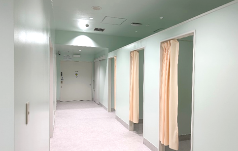 室安静等待约60分钟,直至显像剂药物分布全身。期间不能看手机,不能读书。如果不能保持安静,有可能无法进行检查。
以薄荷绿为基调的舒适空间
拍摄影像
用PET与CT融为一体的设备,拍摄影像。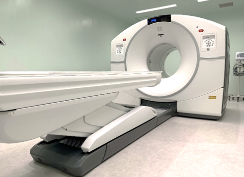
一次拍摄大约需要30分钟左右。
PET/CT装置
(DiscoveryIQ)
等待
拍摄结束后,在休息室等待约30分钟,等到FDG显像剂辐射作用减弱。
结算
所有检查结束后,工作人员会带您到结算窗口,请您在窗口或自动付款及支付费用。
【脑部精检+PET/CT癌症筛查体检】注意事项
| 检查前 | 请仔细阅读「无法接受本体检的人」,如有符合事项,请尽快告知我们。 |
|---|---|
| 从检查前一天到检查结束为止,请避免剧烈运动且不要过度操劳。 | |
| 服用糖尿病药物的患者,请在申请书上填写药物名称。 | |
| 检查当天 | 检查前6个小时需要禁食。只可以饮水(纯净水),带糖分的一切饮料除外。 |
| 糖尿病患者需在检查前6个小时停用糖尿病药物和胰岛素。此外,平时服用的处方药可以照常服用。 | |
| 来院时请不要佩戴任何饰品,并且请穿着容易脱穿的服装。 | |
| 当天请不要化妆,如果手脚上有美甲,有涂指甲油请提前卸掉。美瞳也要取下。 | |
| 体检人不会日语的情况,需要会日语的人陪同做检查。 | |
| 检查后 | 检查结束后,1到2个小时以内请尽量避开人多的地方。 |
| 请尽量多喝水,以便尽快将药物排出体外。 | |
| 检查结束当天,男性也必须坐下排尿。 | |
| 检查结束当天,请避免接触婴幼儿,孕产妇及哺乳期女性。 |
Q&A
| 关于PET/CT(FDG)检查的【Q&A】,请确认PET/CT癌症筛查体检。 | |
| Q | 磁共振检查为什么会产生巨大噪音? |
| A | 为了获取体内器官的位置等信息,需要强大的磁场及发生方向变动的【梯度磁场】。由于高速切换磁场时产生的巨大力量,核磁共振设备内部的线圈会发生震动。因此,根据拍摄方法会产生「哐-哐-」,「哔-哔-」,「哒-哒-哒-」等各种噪音。(摘自日本画像医疗系统工业会策划「核磁共振MRI的Q&A」 |
|---|---|
| Q | 头颅磁共振与头部CT有什么不同? |
| A |
磁共振检查是利用磁性的检查,CT是利用放射线(X线)的检查,并且CT会产生核辐射。 一般来说,磁共振成像的分辨率比CT高,可以更详细地评估脑部的状态。此外,磁共振检查不使用造影剂也能拍摄主要血管的图像。但是磁共振检查无法获得有关骨骼的信息。CT对于检测脑出血,颅骨骨折等发现骨骼异常是有效的。 |
| Q | 磁共振检查时间大约是多少? |
| A | 这次的脑部精检只拍摄头颅,大约需要20分钟。 |
| Q | 检查期间,可以活动身体吗? |
| A | 检查期间请不要活动身体,安静地躺在检查台上。 |
| Q | 检查途中,感到不舒服怎么办? |
| A | 检查前我们会给您呼叫铃,如果感到不适,请按铃告诉我们。 |

